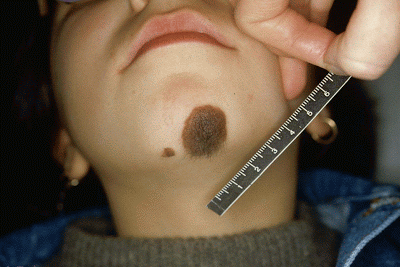
Congenital nevi are visible moles that are usually present at birth and occur as a result of the proliferation of benign melanocytes on the epidermis and dermis.
Article Contents
Congenital Nevus Tardive
This kind of nevi is a matching type to congenital nevi but are usually not present in a newborn at birth. They normally develop during and within the first two years of a child’s life.

Congenital Nevi vs Moles
Developing a mole or a tumor especially on the face is often very alarming for individuals. Therefore, it is of paramount importance to have a distinct explanation of the differences between moles and congenital nevi. Typical moles are widely known as a non-cancerous pigmented tumor while congenital nevi are moles that are present at birth. Babies are not born with moles but develop as they become older.
Types of Congenital Nevi
Speckled lentiginous Nevi

Also referred to as nevus spilus, speckled lentiginous nevus is a kind of congenital melanocytic nevi which is a genetically flat tan mark that is commonly shaped like an oval. The marks are dark spots that form on a flat tan surface. Multiple occurrences of speckled nevus on the skin might be a sign of neurofibromatosis.
Tardive nevi
This kind of nevi appears after birth and grows slower with less amalgamation of the skin melanin compared to congenital nevi. Has an oval shape with an inherited flat tan mark. Multiple formations of tardive nevi may result in neurofibromatosis.
Satellite lesions
Satellite lesions are minor melanocytic lesions that have a small structure. Normally occur on the borders of central congenital melanocytic as they are present in more than 70% of the large congenital nevi. This kind of melanocytic nevi is shaped like an oval and has an inherited flat mark.
Garment nevi
The name garment nevi relate to the anatomical location of the nevus which has an oval shape and a genetically flat tan mark. There are two different kinds of garment nevi as one appears on the entire arm and the shoulders with a coat sleeve while the bathing trunk covers the area around the ass.
Halo phenomenon
This type of congenital melanocytic nevi makes the affected skin area appear light in color with the central lesion smaller and lighter compared to other affected areas. Has an oval shape and a flat tan mark that is inheritable?

Categories of Congenital Nevi
Congenital nevi are normally categorized in respect to their size. All nevi that affect the skin area and should be described concerning their color, body size and surface features. The different classification includes:
- Small congenital melanocytic which are usually less than 1.5 cm in diameter.
- Medium congenital melanocytic which is about 1.5 cm but do not exceed 10 cm in diameter.
- Large congenital nevi which has a diameter of 11-20 cm.
- Giant congenital nevi are above 20 cm -40 cm in diameter.
Congenital melanocytic nevus
Is a multi shaded mark with an oval or round shaped. It has pigmented patches with the skin having increased hair growth. Often where the skin has congenital melanocytic naevi, the skin is rough and bumpy. It usually grows in proportion with the skin; it also increases in size. In the rare case, the mark may disappear or become darker, hairy and bumpier especially around the puberty time.
The frequency of congenital melanocytic nevi appearance
The frequency at which this birthmark is dependent on the different categories of the nevi as small congenital may appear once in every 100 births while medium congenital can appear in 1 of every 1000 births. Of all the different kinds of congenital naevi, the giant melanocytic is less common and appears once in every 20, 000 lives.
This condition is common among all the races, and ethnic groups as males are at risk much like females are.
How sunlight contributes to the development of melanoma
According to research done by the NYU School of medicine overexposure to UV rays is one of the major factors that lead to melanomas. UV rays usually make up a small percentage of the sun rays but are the most distractive effects on the skin as they damage the skin DNA in melanocyte cells. The cancer of the skin is associated with a damaged DNA gene which controls the cell growth.
The development of melanoma is directly attributed to the nature of ultraviolet exposure on the skin. For instances, the development of melanoma on the legs and body trunk which mainly composes of the back and chest is interrelated to repeated sunburns on the skin, especially during childhood. The melanocytes cells contain significant amounts of melanin that usually darkens the skin to protect it from sun rays. These cells are more vulnerable to sun rays compared to normal skin cells.
Most experts are lenient to the fact that in its own context, the sun rays can be attributed to the development of melanoma. The protagonist of this theory argues that there is a significant difference in melanoma on the face and neck compared to ones on the trunk or ones within the internal body surface like the mouth which have low or no sun exposure.
Congenital Nevi Removal
The chances of one developing melanoma are at a high percentage especially within early childhood thus it is important for regular follow up to check whether it may be cancerous. For efficient removal and treatment of congenital melanocytic nevi, these factors should be considered;
- Age of the individual.
- The possibility of an individual developing malicious changes within the lesion.
- The location and depth of the congenital nevi.
- The size of the lesion.
In consideration of the surgical removal, the size of the lesion is important as giant congenital melanocytic should be removed when melanoma has developed within. As for small congenital nevi then the child age is important as well as the size and the growth of the congenital nevi. Surgery should only take place until when the child can cooperate with anesthetic injections.
Congenital Nevi Birthmark Removal
The procedures for safe congenital nevus removal include;
Prophylactic surgical removal
This procedure put into consideration the fact that there is a greater risk of one developing melanoma within the lesion. There is risk involved when performing this procedure such as infection, flap failure, or bleeding.
Laser
Laser method uses ruby Q laser or carbon dioxide resurfacing laser to lighten the surface of the lesion.
Tangential excision
The procedures allow the use of a sharp blade to remove the outermost layer of the skin which comprises of the epidermis and upper dermis. It allows the removal of lesion pigmentation but may not remove the nevi completely. The process of shaving can cause a significant scar.
Chemical peels for congenital nevi treatment
The process, phenol or trichloroacetic acid are used to lighten the pigmentation of the congenital nevus surface which is usually located within the skin upper most layers.
Dermabrasion
This procedure is a partial removal procedure for large congenital nevi as it lightens the color of the nevus. It does not completely remove the hair growth.
Psychological Challenges Related with Congenital Nevi
There is a wide saying that says what does not infect you does not affect you. This common phrase is commonly used in reference to individuals with visible moles or birthmarks such as congenital nevi. These can make one have low self-esteem. Such kind of unwarranted attention can make individuals come across certain psychological issues. These issues include
Outside reaction
Having a largely visible nevus can make one have a fair share of gaze, finger pointing, teasing, murmuring and bullying.
Self-perception
Individuals with congenital nevi can have a negative perception of themselves as they are treated unfairly by others. This leads to one having anxiety issues, low self-esteem and depression issues.
Patient Education
It is important that one understand that having a visible nevus is normally and patients should learn to embrace it for prosperity. With surgical methods, one can remove the congenital nevi so patients should not feel embarrassed.
Congenital Nevi Pictures


I had a mole removed like a month or 2 ago and the doctor said it was permal nevus. So he removed it and then the results came back and he said that it was permal nevus i don’t no what that means but he said that its possible that sometimes cancer can be located not in the mole but in the skin where the mole was now he didn’t say i have cancer but he said he wanted to take more just to make sure so i said i wanted to go to a dermatoligist and he said there isent anything to worry about there is no reason for him to take of more skin but i am still worried that i needed more off??? what does permal nevus mean? i know what beign means but idk i am scared still!
Hi Audie. I assume you meant to say dermal nevus. Dermal nevi are common benign and tend to occur more in light skinned individuals. They are simply an increase in the number of cell on the layer of skin just below the epidermis. They tend to increase in number with age up until thirties when they begin to regress with age. Dermal nevi also change morphology as they grow deeper into the dermis.
It is okay for you to worry. Kindly consult with your dermatologist. I really hope this helps.